Note that your final mark will not be saved in the system.
3.1.1.5 The musculo-skeletal system and analysis of movement Typeit
Type the correct answers into the spaces. Fill all the spaces before clicking ‘Check Answers!’
Skeletal muscles work in pairs to cause movement at the joints they articulate. As one muscle or muscle group in the pair contracts, the other relaxes, working in what is called an antagonistic pair. The muscle responsible for pulling on the joint to cause movement is termed the , while the paired muscle that is passive during the movement is known as the .
As well as the different roles muscles play in movement, there are a variety of contraction types that affect how movement is performed:
- muscle contractions are characterised by a change in the length of the muscle. This type of contraction can be further divided as:
- (i.e. the lengthening of the muscle) and
- (i.e. the shortening of the muscle) muscle contractions.
- Muscle contraction that occurs while there is no change in the muscle’s length is known as an contraction.
When a basketball player jumps to perform a slam dunk, the gastrocnemius acts as the muscle, causing plantar flexion at the ankle joint, allowing the player to leap into the air. The gastrocnemius shortens as it contracts; this is an muscle contraction. At the same time, the tibialis anterior works as the muscle by relaxing to allow plantar flexion to be performed. When the athlete lands, the gastrocnemius lengthens as it contracts, producing tension to control the dorsiflexion movement. This is known as an isotonic contraction.
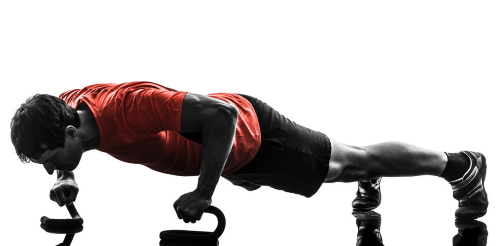
During a press-up, holding a static position during the downward phase (as shown in the image above) is an example of an muscle contraction at the triceps brachii, which remains under tension to maintain a stable horizontal body position.
Examples of other antagonistic muscle pairs in the body include the following:
| Joint | Joint action | Agonist(s) | Antagonist(s) |
| Shoulder | Flexion | Anterior deltoid | Latissimus dorsi, teres minor and posterior deltoid |
| Extension | Latissimus dorsi, teres minor and posterior deltoid | Anterior deltoid | |
| Abduction | Middle deltoid | ||
| Adduction | Latissimus dorsi | Middle deltoid | |
| Horizontal abduction | Trapezius, posterior deltoid and teres minor | Pectoralis major, anterior deltoid | |
| Horizontal adduction | and anterior deltoid | Trapezius, posterior deltoid and teres minor | |
| Hip | Flexion | Iliopsoas and hip – rectus femoris, iliacus, psoas, iliocapsularis and sartorius | Gluteals |
| Extension | Gluteals | Iliopsoas and hip flexors –rectus femoris, iliacus, psoas, iliocapsularis and sartorius | |
| Abduction | Tensor fascia latae and gluteus medius and minimus | Hip adductors – adductor brevis, longus and magnus | |
| Adduction | Hip adductors – adductor brevis, longus and magnus | Tensor fascia latae and gluteus medius and minimus | |
| Horizontal abduction | Tensor fascia latae and gluteus medius and minimus | Hip adductors –adductor brevis, longus and magnus | |
| Horizontal adduction | Hip adductors – adductor brevis, longus and magnus | Tensor fascia latae and gluteus medius and minimus | |
| Elbow | Flexion | Biceps brachii | Triceps brachii |
| Extension | Triceps brachii | Biceps brachii | |
| Knee | Flexion | Hamstring group – semitendinosus, semimembranosus and biceps femoris | Quadriceps group – rectus femoris, vastus lateralis, vastus medialis and vastus intermedius |
| Extension | Quadriceps group – rectus femoris, vastus lateralis, vastus medialis, vastus intermedius | Hamstring group – semitendinosus, semimembranosus, and biceps femoris |